While researchers often focus on CD8+ T cells in antitumor immunity, many studies have shown critical roles for CD4+ T cell responses as well. In order to better understand the context in which antitumor CD4+ T cell responses are initiated, Binnewies and Mujal et al. examined a variety of cell types and factors in tumors and tumor-draining lymph nodes (tdLNs) and explored possible therapeutic interventions to enhance the magnitude and quality of CD4+ T helper 1-like responses in both mice and humans.
Binnewies and Mujal et al. began by using single-cell RNAseq (scRNAseq) to characterize myeloid cells from tdLNs from B16F10 melanoma-bearing mice. From this data, they identified multiple clusters of both resident and migratory conventional dendritic cells (cDCs). In particular, they focused on two clusters, which bore a migratory cDC2 signature (Zbtb46, FLT3) and could be distinguished as either CD301b+ or CD301b-. The CD301b+ cDC2 subset expressed markers associated with cells of a monocyte/macrophage lineage and high levels of inhibitory receptors such as PD-L2. In a separate in vivo experiment, both subsets showed the capacity to bear tumor antigen and migrate in a CCR7-dependent manner from the tumor to the tdLN. These migratory cDC2 populations were found to be essential for the proliferation of transferred tumor-specific CD4+ T cells (OVA-specific OT-II cells). Ex vivo coculturing of either cDC2 subset with OT-II cells induced OT-II expansion, suggesting that the cDC2 subsets have overlapping function as inducers of antitumoral CD4+ T cell priming.
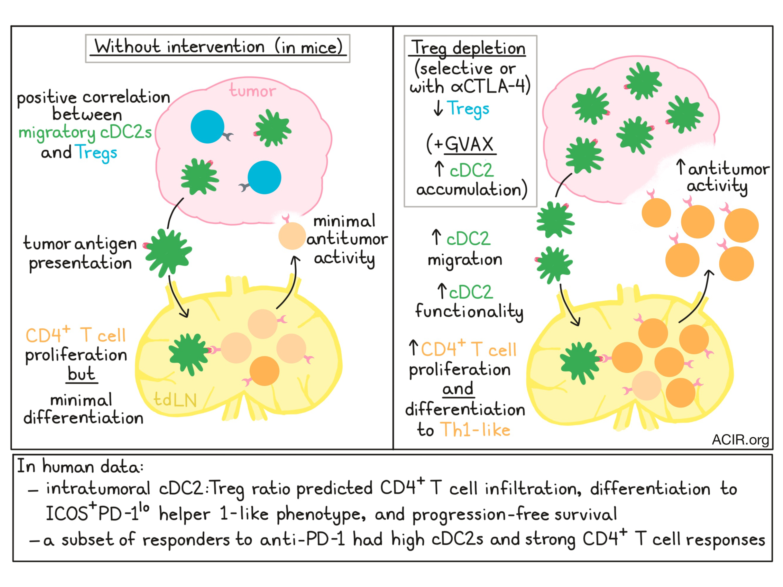
Despite the evidence that migratory cDC2s prime antitumor CD4+ T cells, antitumor immunity was not evident in tumor-bearing mice. To determine whether CD4+ T cells were properly differentiating into effector cells, the researchers compared the activation of OT-II cells in the tumor-draining lymph node to that in a tolerizing LN (induced by OVA injection without adjuvant) or in the context of an inflammation-inducing virus. Compared to the viral setting, activation of OT-II cells in the tumor-draining lymph node or tolerizing lymph node was low. Differentiation of CD4+ T cells towards the ICOS+PD-1lo Th1 phenotype (associated with antitumor response) and cytokine production upon restimulation were also minimal in both of the non-inflamed lymph node settings, indicating that CD4+ T cell differentiation was in some way defective.
Based on prior evidence that expansion of cDC2s does not result in the expansion of conventional CD4+ T cells, the researchers hypothesized that cDC2 expansion may instead induce expansion of Tregs as a feedback mechanism to inhibit the cDC2 population. Supporting this hypothesis, the team found a correlation between the frequency of cDC2s and the frequency of Tregs in tumors. Selective depletion of Tregs induced tumor rejection, dependent on the migration of cDC2 cells to the tdLN, the presentation of tumor antigens by cDC2s, priming of CD4+ T cells in the tdLN, and egress of CD4+ T cells from the tdLN. OT-II cells transferred into tumor-bearing, Treg-depleted mice showed increased expansion and increased expression of activation markers. ScRNAseq showed that following Treg depletion, cDC2s increased the expression of genes related to costimulation, T cell chemoattraction, and response to pro-inflammatory cytokines. This coincided with profound increases in ICOS+PD-1lo Th1-like CD4+ T cells in the tdLN, further supporting the hypothesis that Tregs suppress the ability of cDC2s to effectively prime CD4+ conventional T cells.
Because anti-CTLA-4-based therapies may induce antitumor immunity by depleting Tregs, Binnewies and Mujal et al. reasoned that they may also work to enhance the functionality of cDC2s. In mice, the use of an anti-CTLA-4 clone to selectively deplete Tregs from the tumor, but not the tdLN, led to increased migration of CD301b+ cDC2s to the tdLN, increased functionality of cDC2s, and a significant increase in ICOS+PD-1loCD4+ conventional T cells in both the tdLN and tumor, confirming that Treg depletion in the context of anti-CTLA-4 enhances cDC2 migration and function.
As GM-CSF has previously been shown to induce cDC2 expansion, Binnewies and Mujal et al. tested the combination of GVAX with anti-CTLA-4. Alone, GVAX increased the numbers of both cDC2 subsets and enhanced the functionality of the CD301b+ cDC2 subset. The combination of GVAX and anti-CTLA-4 reduced Tregs, increased the numbers and enhanced the functionality of both cDC2 subsets, induced robust expansion of CD4+ T cells, and reduced tumor growth.
To better understand whether their observations in mice would be relevant in humans, the researchers evaluated scRNAseq data from myeloid cells derived from a human melanoma-draining lymph node. They observed heterogeneity in the human migratory cDC2 subset, marked by expression of BDCA-1, that closely resembled the heterogeneity observed in mice. The data from mice had suggested that the intratumoral densities of cDC2s and Tregs could predict the quantity and quality of CD4+ T cells. To determine whether these markers had similar predictive value in humans, the researchers analyzed 32 primary tumors from the head and neck region (typically rich in Tregs), and defined three TME types. Patients with low levels of cDC2s in the primary tumor, or with high levels of cDC2s and high levels of Tregs, demonstrated low levels of CD4+ T cell infiltration. Patients with high levels of cDC2s and low levels of Tregs, however, showed more robust CD4+ T cell infiltration – many of which were ICOShiPD-1lo – and longer progression-free survival. To determine whether the presence of cDC2s might serve as a biomarker in the context of PD-1 blockade, the researchers analyzed data from a prior study and found that responders to anti-PD-1 could be divided into two categories: those with high BDCA-3+ cDC1s and strong CD8+ T cell responses, or those with high BDCA-1+ cDC2s and strong CD4+ T cells responses. It is therefore possible that the presence of cDC2s could serve as an additional biomarker for a subset of patients likely to respond to checkpoint blockade.
by Lauren Hitchings
Meet the Researcher
This week, we interviewed Mikhail Binnewies and Adriana Mujal, first co-authors on this paper, as well as Max Krummel, the lead author.

What prompted you to tackle this research question?
MB: An efficacious adaptive T cell response is critical for productive antitumor immunity. Recent data, however, suggests that in certain patients, T cells present within the tumor are terminally exhausted and unlikely to be revived by checkpoint inhibitors. Our group believes that by understanding how antitumor T cell responses are initiated, we can find ways to boost the tumor infiltration of newly activated antitumor T cells. Central to T cell activation are dendritic cells, an innate immune population that functions to survey tissues for pathogens or tumor and guide adaptive immune responses based on what it has encountered.
AM: We were also interested in better understanding settings in which CD4+ T cells are key contributors to antitumor immunity and how these T cell populations could be utilized or leveraged to achieve productive antitumor responses in certain cohorts of patients.
What was the most surprising finding of this study for you?
MB: What surprised us the most was that the mechanism of immune activation and suppression we identified in mouse models was so closely replicated in human patients. To us this indicates that in certain tumor indications, targeting this suppressive interaction therapeutically could be a potent way to boost antitumor immunity.
AM: I always enjoy the surprise of the subsequent questions that emerge from one's original question and findings. For example, what mechanisms give rise to the heterogeneity in cellular proportions within the patient tumors? Why and how do pathways of immune suppression act predominantly on certain arms of the immune system?
What was the coolest thing you’ve learned (about) recently outside of the lab?
MB: Classical conditioning in dog training. My wife and I recently adopted a sweet but very shy dog. It is amazing how much you can change a dog’s reaction to triggering objects or environments by associating it with something they enjoy. Pavlov was right!
AM: Central Park has over 275 species of birds. Having moved to NYC about a year ago, I've been enjoying ways to see the city and appreciate elements that I would have previously not noticed.
What prompted you to tackle this research question?
While a large cohort of immune responses to cancer take place using CD8+ T cells, we had been finding archetypal classes of patients whose immune responses were more dominated by CD4+ T cells. We wanted to understand what was keeping these CD4+ T cells from destroying the tumor.
What was the most surprising finding of this study for you?
We found three types of patients that exactly phenocopy what we’d seen in mouse models. And the outcomes in mouse models completely predicted the outcomes in humans. It’s not surprising in the long run, but mouse models can very much predict human biology – it would just seem to be the case of figuring out what parameters are the most important for defining classes of patients and then matching them.
What was the coolest thing you’ve learned (about) recently outside of the lab?
Outside my lab but in our field: Non-cancerous somatic mosaicism – that as our genomes succumb to mutation (entropy) that some variants ‘win’ and things like our skin and our epithelium become a mosaic of colonies, like players on a risk board. Some presumably then ‘win big’ and become too good at proliferation in tissue and become cancer. But our immune systems must deal with huge amounts of variation in ‘self’ proteins, regardless of cancer.
Outside of our field: The albatross can migrate for over 1200 miles, then stops for a feed when it finds krill and can eat so much it cannot take off to fly again until it loses some weight.


